
Body Fluids and Circulation
Structure of human heart and blood vessels
- Harvey discovered circulatory system.
- Human heart is located between the lungs in the thoracic cavity where it weighs from 280-340g in male, and 230-280 gm in female.
- Pericardium is a protective covering consisting of outer parietal pericardium and inner visceral pericardium. where in between is filled with pericardial fluid that allows free movements of heart.
- The mammalian heart comprises of 4 complete chambers
- The right atrium receives deoxygenated blood from superior vena cava (SVC), inferior vena cava (IVC) and coronary sinus. The left atrium which is thicker than right ventricle receive oxygenated blood from pulmonary (2 lungs) through 4 pulmonary vein.
- Heart is of two types (i) myogenic heart (eg: mammals, mollusca) (ii) neurogenic heart (annelida, arthropoda)
- A patch of modified heart muscle (myogenic heart) is present in the wall of right atrium called as SA node (sinoatrial) (@Ka Pace maker of heart).
- Another aggregate of modified heart muscle tissue called as AV node (Atrioventricular) is present at the lower right and of interatrial septum.
- A mass of specialised fibers, the bundle of His, originates from AV node which divides into two branches one going to each ventricle.
- Klithin the myocardium of ventricles the branches of bundle of his divide into network of fire fibers, called purkije fibers. Which convey impulse of contraction from AV nodes to ventricles.
- Superior vena cava also called as precaval system is guarded by haversian valve whereas inferior vena cava (post caval system) is graded by eustachian valve.
- The left atrium is less in volume than that of right atrium but is has thicker walls.
- The pulmonary trunk arises from right ventricle which divides into left and right pulmonary arteries that carry deoxygenated blood to lungs.
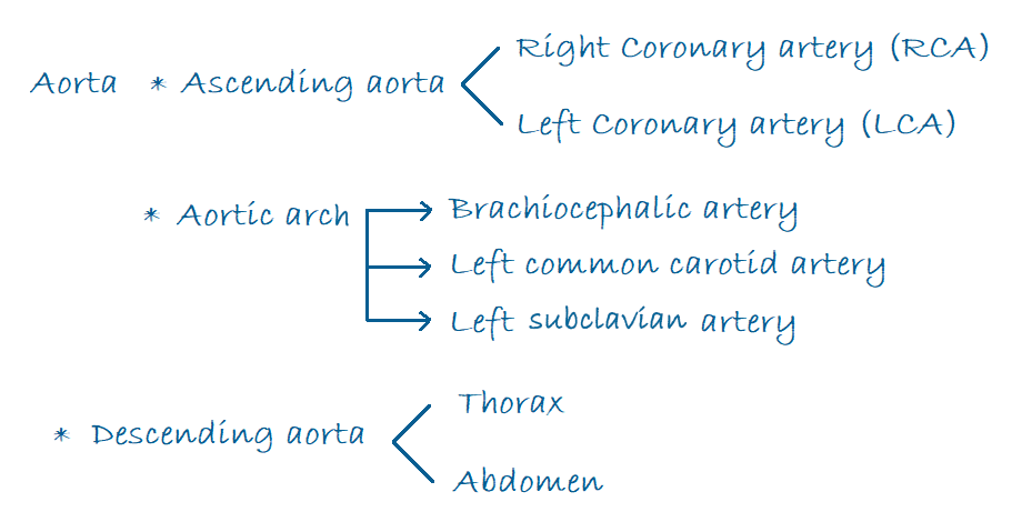
- The aorta arises from left ventricle which is divisible into ascending aorta, aortic are and descending aorta.
- Left atrium and left ventricle is guarded by Bicuspid valve (mitral valve) which has two flaps, and the right atrioventricular opening is guarded by tricuspid valve.
- Attached to both the walls are special fibrous cords called "chordae tendiae" which are joined to the other ends with special muscles called papillary muscles.
- At the base of pulmonary trunk and aorta are located pulmonary and aortic semilunar valve.
Watch this video for the topic Structure of human heart and blood vessels from 0:48 to 30:14
Disclaimer: Compete.etutor.co may from time to time provide links to third party Internet sites under their respective fair use policy and it may from time to time provide materials from such third parties on this website. These third party sites and any third party materials are provided for viewers convenience and for non-commercial educational purpose only. Compete does not operate or control in any respect any information, products or services available on these third party sites. Compete.etutor.co makes no representations whatsoever concerning the content of these sites and the fact that compete.etutor.co has provided a link to such sites is NOT an endorsement, authorization, sponsorship, or affiliation by compete.etutor.co with respect to such sites, its services, the products displayed, its owners, or its providers.